Xiralite Deutschland
 Xiralite United States

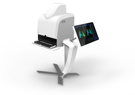
Was ist Xiralite?
Ein schnelles, innovatives und reproduzierbares Diagnoseverfahren zur strahlungsfreien Untersuchung von bis zu 30 Gelenken in beiden Händen bei unterschiedlichen Erkrankungen zur Unterstützung klinischer Entscheidungen in der Praxisroutine.
Mehr erfahren